
Basic Anatomy of the Human Heart Cardiology Associates of Michigan
Heart. Your heart is the main organ of your cardiovascular system, a network of blood vessels that pumps blood throughout your body. It also works with other body systems to control your heart rate and blood pressure. Your family history, personal health history and lifestyle all affect how well your heart works.
Anatomical Drawing Heart at GetDrawings Free download
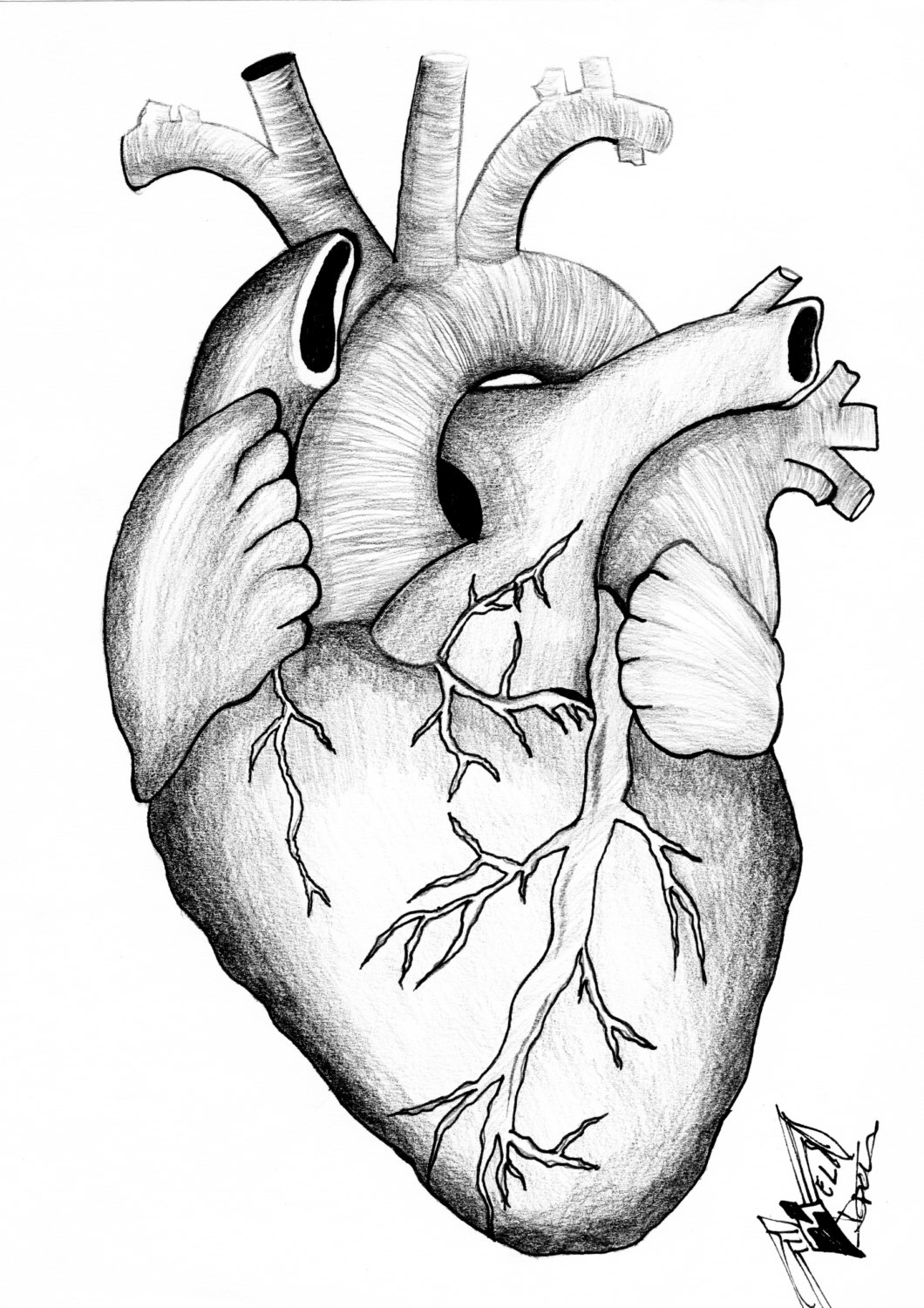
1 Draw a tilted and irregular curved shape in the center of your page. Use a pen or pencil to draw the heart's main body. Create a curved shape similar to an acorn or apple's bottom half. Angle the slightly tampered end of the shape to the left about 120 degrees. [1] The main shape will be the basis for the left and right ventricles.

Heart Anatomy chambers, valves and vessels Anatomy & Physiology
161 Share 18K views 3 years ago Body Part Drawings: How to Draw Body Parts With this easy human heart drawing ideas, you can learn how to draw a human heart easily. I made this cool.
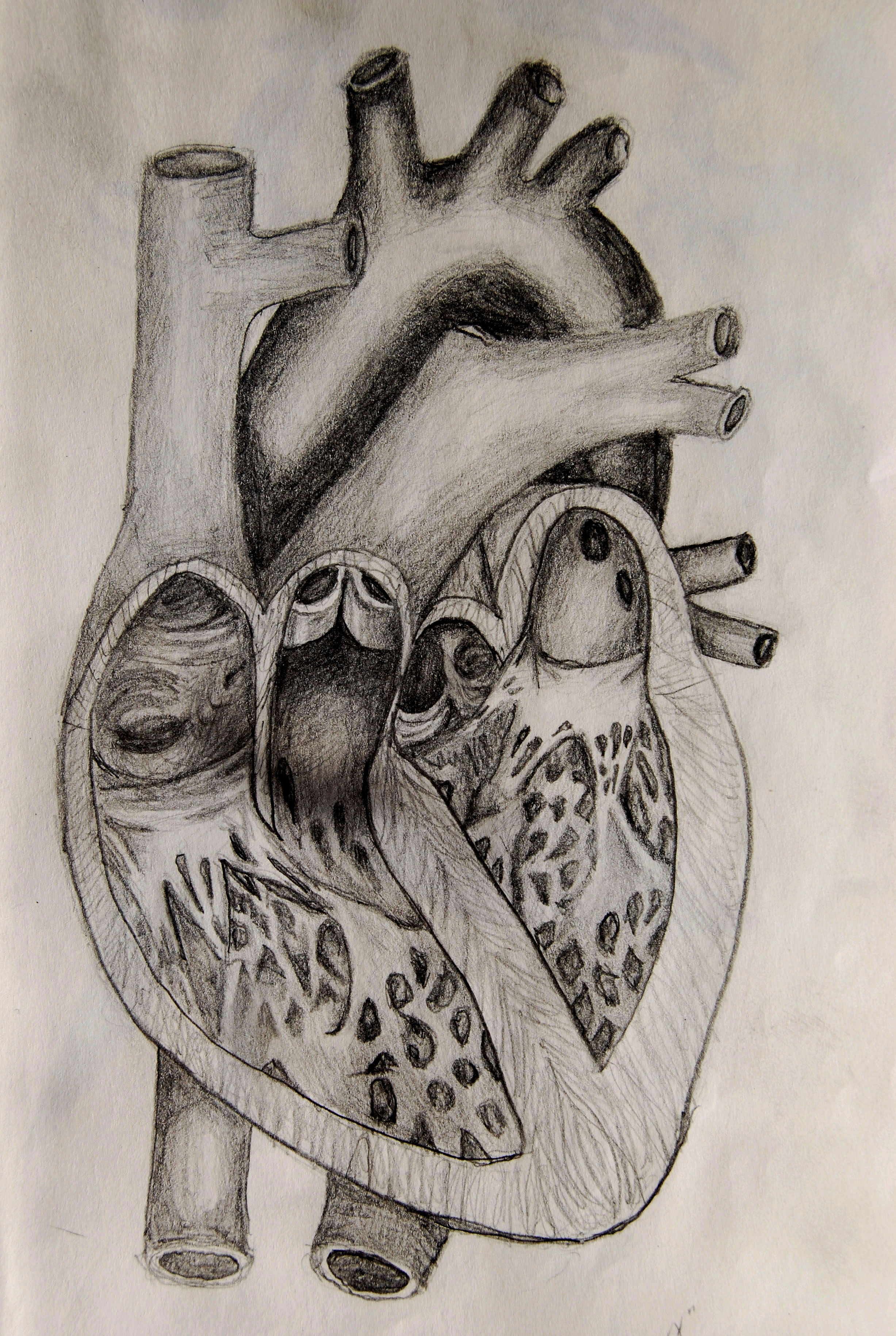
Realistic Human Heart Drawing at GetDrawings Free download
$9.99 Add To Cart Anatomy of the Heart Welcome to the anatomy of the heart made easy! We will use labeled diagrams and pictures to learn the main cardiac structures and related vascular system. In addition to reviewing the human heart anatomy, we will also discuss the function and order in which blood flows through the heart.
Anatomical Drawing Of The Heart at GetDrawings Free download
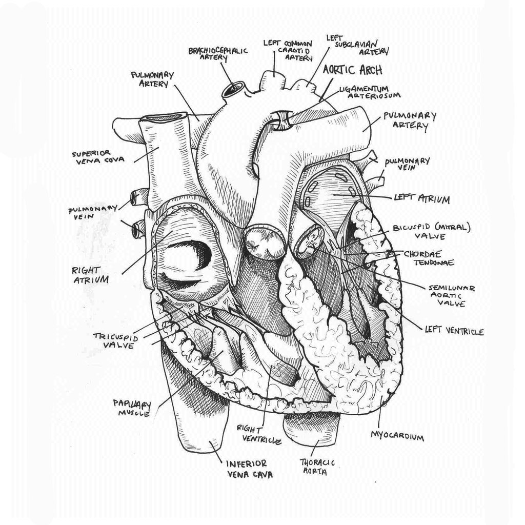
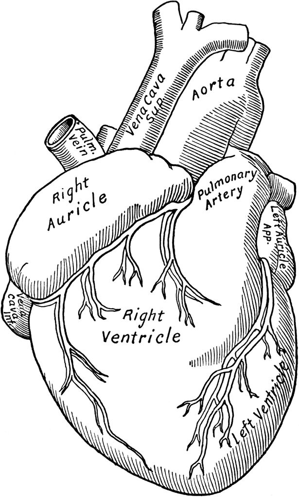
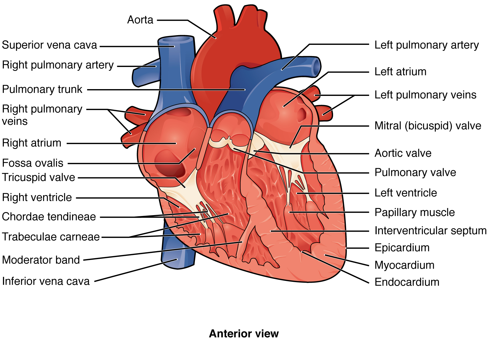
Interactive quizzes Sources + Show all Labeled heart diagrams Take a look at our labeled heart diagrams (see below) to get an overview of all of the parts of the heart. Once you're feeling confident, you can test yourself using the unlabeled diagrams of the parts of the heart below. Labeled heart diagram showing the heart from anterior
Human Heart Line Drawing at GetDrawings Free download
In this short time-lapse video I show how to draw the anatomical human heart in a simplistic way, showing the various parts and how they relate to each other.

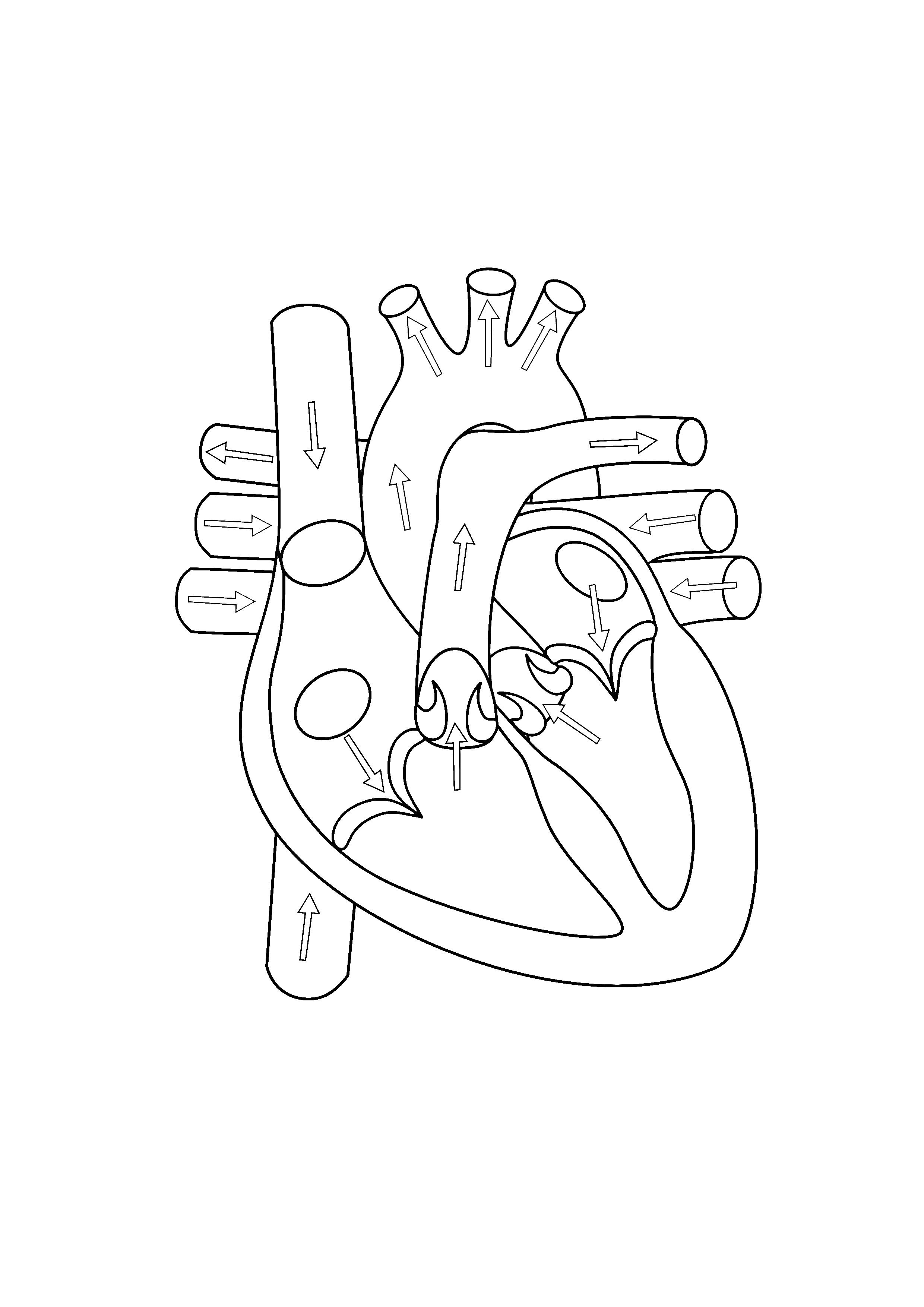
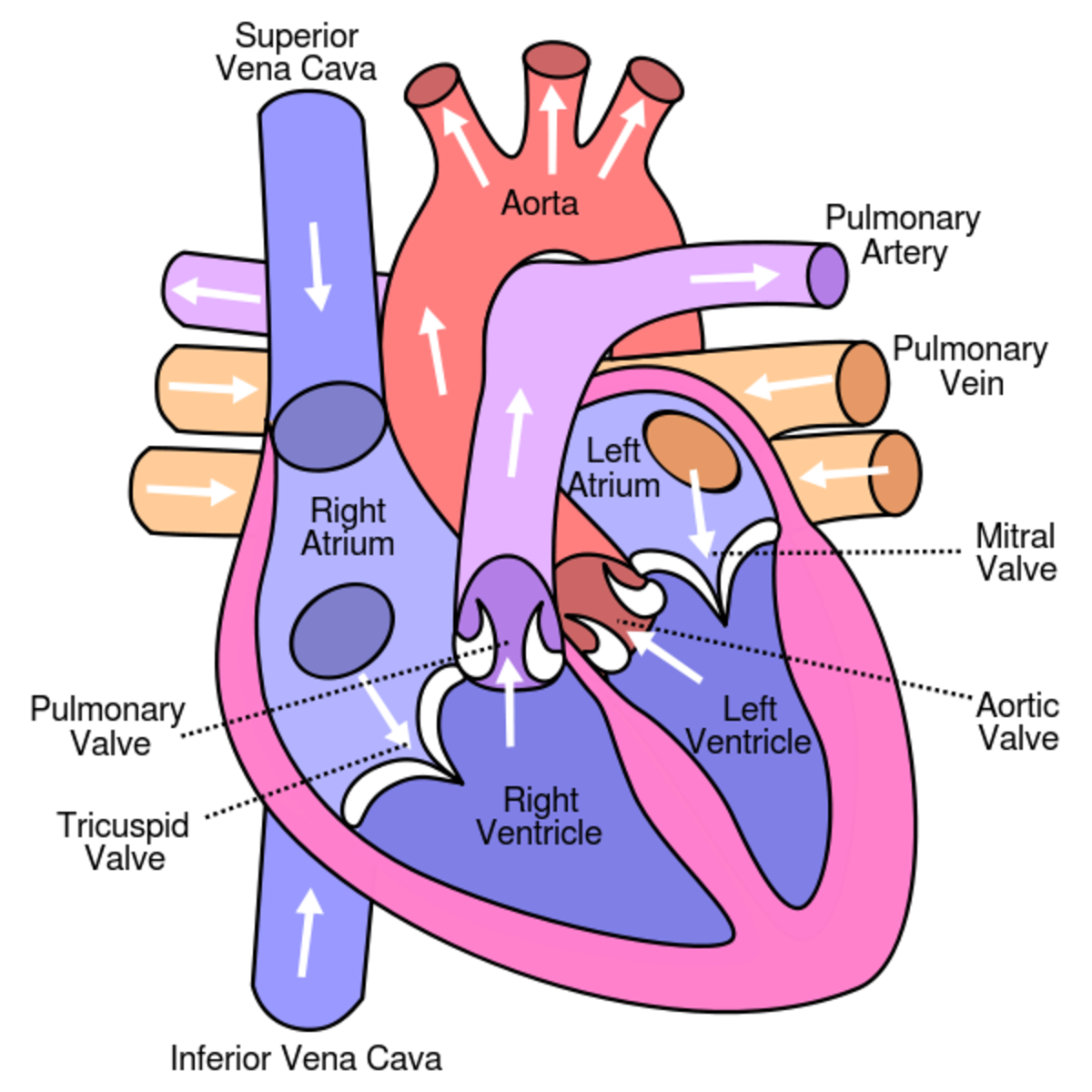
Draw a heart. label the chambers and draw arrows to show the flow of
1 To find a good diagram, go to Google Images, and type in "The Internal Structure of the Human Heart". Find an image that displays the entire heart, and click on it to enlarge it. [1] 2 Find a piece of paper and something to draw with. Start with the pulmonary veins. They will be to the lower left of the Aorta. There are two of them.
Heart Anatomy · Anatomy and Physiology
Easy Human Heart Drawing - Step 5. 5. Next, draw the right atrium and the superior vena cava. For the atrium, enclose an irregular form at the junction of the ventricle and the aortic arch. Above this, draw a narrow vertical oval along the arch, and connect this to the atrium using a curved line.

Cardiovascular Disease
In this lecture, Dr Mike shows the two best ways to draw and label the heart!

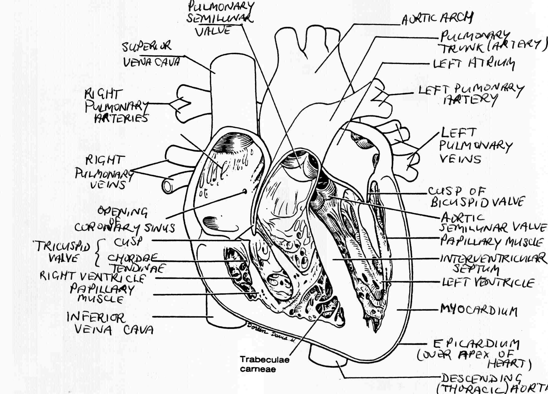
How to Draw the Internal Structure of the Heart 13 Steps
heart anatomy drawing illustrations and vector graphics available royalty-free, or start a new search to explore more great images and vector art. xxxl very detailed human heart - heart anatomy drawing stock illustrations

31 Human Heart To Label Labels Design Ideas 2020
Drawing Internal Anatomy of the Heart. A drawing of the anatomy of the opened normal heart, with English labels. From the OpenStax Anatomy and Physiology book. Valves. Video Beating Aortic Valve. A video of the aortic valve in a cadaver heart that is beating in a laboratory set-up by the University of Minnesota.
Human Heart Anatomy Drawing at GetDrawings Free download
Exercise regularly Quit smoking Manage stress What should I expect during a cardiology appointment at Nao Medical? During your appointment, our cardiology specialists will perform a thorough evaluation of your heart health, which may include a physical exam, diagnostic tests, and a review of your medical history.

How to Draw the Internal Structure of the Heart (with Pictures)
The heart is a muscular organ that pumps blood around the body by circulating it through the circulatory/vascular system. It is found in the middle mediastinum, wrapped in a two-layered serous sac called the pericardium.

Heart Diagram Sketch at Explore collection of
This interactive atlas of human heart anatomy is based on medical illustrations and cadaver photography. The user can show or hide the anatomical labels which provide a useful tool to create illustrations perfectly adapted for teaching. Anatomy of the heart: anatomical illustrations and structures, 3D model and photographs of dissection
Learn About the Heart and Circulatory System for Kids HubPages
Location of the Heart. The human heart is located within the thoracic cavity, medially between the lungs in the space known as the mediastinum. Figure 19.2 shows the position of the heart within the thoracic cavity. Within the mediastinum, the heart is separated from the other mediastinal structures by a tough membrane known as the pericardium.
29+ Draw The Diagram Of Human Heart Images Diagram 123
In animals with lungs —amphibians, reptiles, birds, and mammals—the heart shows various stages of evolution from a single to a double pump that circulates blood (1) to the lungs and (2) to the body as a whole. In humans and other mammals and in birds, the heart is a four-chambered double pump that is the centre of the circulatory system.