
Parts Parts Of A Microscope
With Labeled Diagram and Functions How does a Compound Microscope Work? Before exploring microscope parts and functions, you should probably understand that the compound light microscope is more complicated than just a microscope with more than one lens.

Microscope, Microscope Parts, Labeled Diagram, and Functions
What are the optical parts? There are two types of optical microscopes and these are the simple and compound microscopes. There are various types of microscopes and each type has a specific set of functions. The common types of microscopes are: Simple microscope - It was the first microscope ever created.
Parts of a Microscope The Comprehensive Guide Microscope and
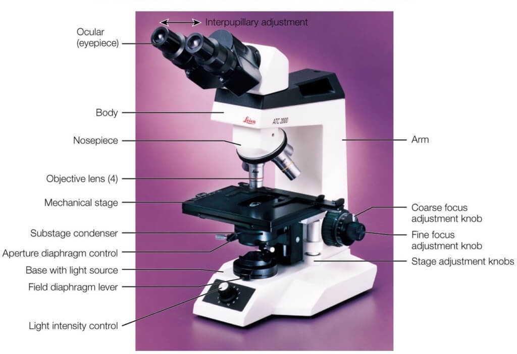
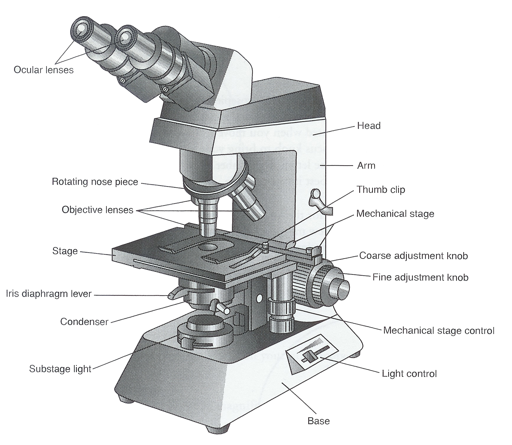
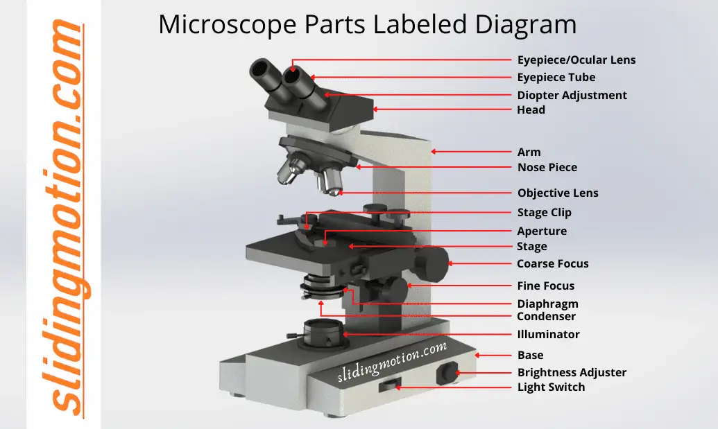
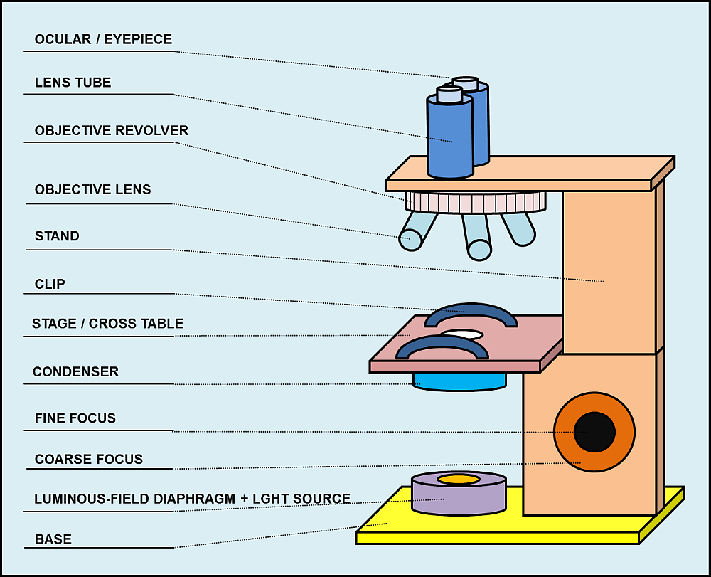
Structural parts of a microscope: There are three major structural parts of a microscope. The head comprises the top portion of the microscope, which contains the most important optical components, and the eyepiece tube.; The base serves as the microscope's support and holds the illuminator.; The arm is the component of the microscope that connects the eyepiece tube to the base of the.
Anatomy Of A Microscope
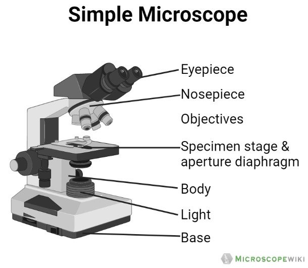
Simple microscope is a magnification apparatus that uses a combination of double convex lens to form an enlarged, erect image of a specimen. The working principle of a simple microscope is that when a lens is held close to the eye, a virtual, magnified and erect image of a specimen is formed at the least possible distance from which a human eye.
Parts of a Microscope Labeling Activity
All microscopes share features in common. In this interactive, you can label the different parts of a microscope. Use this with the Microscope parts activity to help students identify and label the main parts of a microscope and then describe their functions.. Drag and drop the text labels onto the microscope diagram. If you want to redo an answer, click on the box and the answer will go back.

Labeled Microscope Diagram Tim's Printables
The microscope illustrated in Figure 5 below was manufactured by Hugh Powell and Peter Lealand around 1850. The tripod base provided a sturdy support for the microscope, which many people consider the most advanced of its period. Parts of a Powell and Leland Microscope Diagram
Parts Parts And Functions Of A Microscope
The hand magnifying glass can magnify about 3 to 20×. Single-lensed simple microscopes can magnify up to 300×—and are capable of revealing bacteria —while compound microscopes can magnify up to 2,000×. A simple microscope can resolve below 1 micrometre (μm; one millionth of a metre); a compound microscope can resolve down to about 0.2 μm.
Parts of the Microscope (Labeled Diagrams) Simple and Compound Microscope
There are 1000 millimeters (mm) in one meter. 1 mm = 10 -3 meter. There are 1000 micrometers (microns, or µm) in one millimeter. 1 µm = 10 -6 meter. There are 1000 nanometers in one micrometer. 1 nm = 10 -9 meter. Figure 1: Resolving Power of Microscopes. The microscope is one of the microbiologist's greatest tools.
Guide to understand microscope parts, names, functions & diagram
Major structural parts of a compound microscope. There are three major structural parts of a compound microscope. The head includes the upper part of the microscope, which houses the most critical optical components, and the eyepiece tube of the microscope.; The base acts as the foundation of microscopes and houses the illuminator.; The arm connects between the base and the head parts.
Parts Of A Microscope With Functions And Labeled Diagram Images
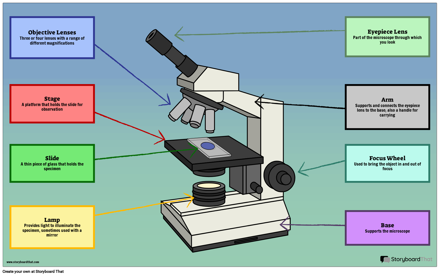
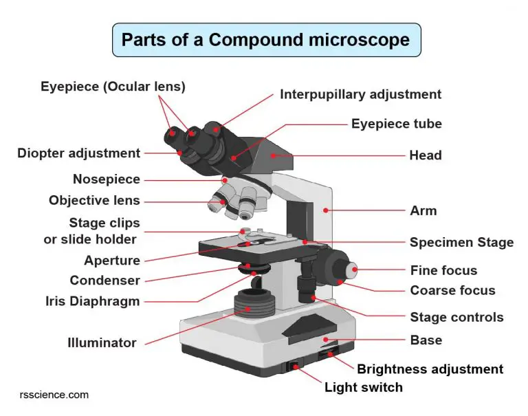
The individual parts of a compound microscope can vary heavily depending on the configuration & applications that the scope is being used for. Common compound microscope parts include: Compound Microscope Definitions for Labels Eyepiece (ocular lens) with or without Pointer: The part that is looked through at the top of the compound microscope.

🎉 Main components of a light microscope. Parts of a microscope with
Magnification is a measure of how much larger a microscope (or set of lenses within a microscope) causes an object to appear. For instance, the light microscopes typically used in high schools and colleges magnify up to about 400 times actual size. So, something that was 1 mm wide in real life would be 400 mm wide in the microscope image.
15 Microscope Parts A Guide on their Location and Function
Explore the different parts of a microscope using a diagram, including the microscope lens, eyepiece, and stage. Updated: 10/13/2022 What is a Microscope? A microscope is a scientific.

Parts of a microscope with functions and labeled diagram
1. Head (Body) The head, also referred to as the body of the microscope, is a structural component that contains the optical parts of the microscope. The figure below shows the area of the microscope considered to be the body of the microscope.
The Wonders Of Microscopes What You Need To Know Creyentes Diverses News
A labeled diagram of microscope parts furnishes comprehensive information regarding their composition and spatial arrangement within the microscope, enabling researchers to comprehend their function effectively. In this comprehensive article, we will delve into the intricate parts of the microscope, exploring their functions in detail.
Compound Microscope Parts Labeled Diagram and their Functions Rs
A light microscope is a biology laboratory instrument or tool, that uses visible light to detect and magnify very small objects and enlarge them. They use lenses to focus light on the specimen, magnifying it thus producing an image. The specimen is normally placed close to the microscopic lens.
Parts and components of light microscopes Light Microscope
2. Compound Microscope. Compound Microscope is a type of microscope that used visible light for illumination and multiple lenses system for magnification of specimen. Generally, it consists of two lenses; objective lens and ocular lens. It can magnify images up to 1000X.
